When a doctor determines a patient has a brain tumor, the next step is usually surgery. Knife in hand, the surgeon is soon presented with an inevitable conundrum: how much is the right amount to cut away? Too little and, if the tumor is malignant, the patient will be subjected to more toxic chemotherapy or radiation than necessary. Too much, and the doctor will be slicing into healthy tissue—and possibly causing the patient undue brain damage.
Yet this high-stakes surgical process can be surprisingly rudimentary. “Surgeons don’t have a very good way of knowing when they’re done cutting out a tumor,” Jonathan Liu, a University of Washington assistant professor of mechanical engineering, said in a press release. “They’re using their sense of sight, their sense of touch, pre-operative images of the brain—and oftentimes it’s pretty subjective.”
Which is why research groups and companies around the world have been working to build technology that can distinguish between cancerous and non-cancerous cells, right in the operating room. A device developed by Liu’s UW research group, along with partners at the Memorial Sloan Kettering Cancer Center, Stanford University and the Barrow Neurological Institute, is on the frontlines.
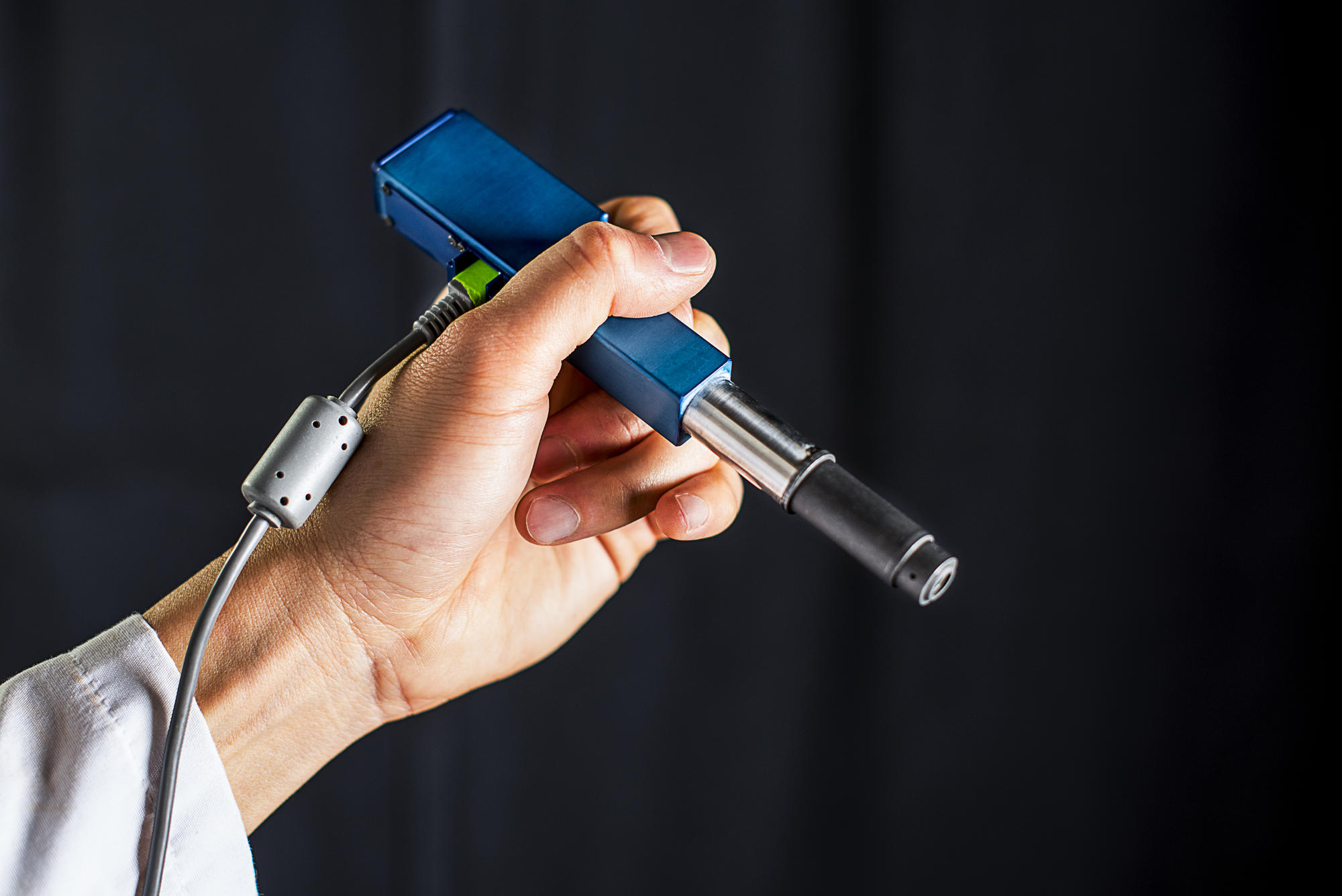
The microscope, described in a paper published in Biomedical Optics Express in January, is about the size of a pen, and can quickly generate images of the tissue in question at the cellular level. It operates via “dual-axis confocal microscopy,” which means it uses a pinpoint of light to illuminate the tissue, and then the light travels down a separate collection path. This technology allows researchers to optically section through tissues, Liu explained in a video. “In other words, we’re able to visualize thin sections of tissues without having to cut it with a knife. We can actually use a light to cut through the tissues and visualize it in three dimensions.”
This could enable doctors to tell which cells to remove—and which to let be—while surgery is underway.
The new microscope’s ability to pinpoint abnormal cells could have another significant application: replacing biopsies. “Typically the only way doctors can really know if it’s cancer or not is to biopsy the tissue,” Liu, who has been working on this technology since 2010, told Crosscut.
This involves a doctor removing a sample of the person’s organ and sending it to the lab, where a pathologist prepares a slide to analyze the tissue by microscope. Not only is the process of taking the tissue invasive, the lag between extracting the sample and giving the patient his or her results can be several days—a long time to have the uncertainty of a threat like cancer weigh on you.
But the handheld microscope could noninvasively discover cancerous cells that have formed in certain organs, as it can provide images of tissue up to half a millimeter below the surface. That doesn’t sound like a lot but, according to Liu, most cancers start at the surface and then spread inward. This is especially pertinent to skin or mouth cancers: with a microscope like this, doctors and dentists could routinely and easily scan these organs, which could help diagnose cancers that form there much earlier on.
“There are a lot of lesions in our mouths that look suspicious,” Liu explained. But most of them turn out to be non-threatening, like canker sores. “They don’t want to biopsy hundreds of tissues that are most likely normal.”
So instead, dentists and oral surgeons usually wait to biopsy it until it looks really bad—by which point, if it is cancer, it has become much more difficult to treat.
The new microscope, which was funded by two grants totaling in $4.1 million from the National Institute of Health, isn’t the only one of its kind. In some ways the UW group is already behind. Caliber I.D. (formerly known as Lucid Technologies, Inc.), a medical technologies company based in Andover, Mass., came out with the handheld VivaScope 3000 in 2013, and Mauna Kea Technologies, based Paris, got FDA clearance to use their Cellvizio in the U.S. last October.
Still, Liu thinks the new microscope is up to the competition. “They all have certain tradeoffs in performance,” Liu said. He explained that their device is smaller than Caliber I.D.’s, which would make it better suited for brain tumor surgeries and easier to fit into patient’s mouths to detect oral cancer, and it provides more detailed images than Mauna Kea’s.
The UW researchers are currently in the process of assembling a second microscope, and they want to put one of them into patient use for the first time later this year (so far, their primary subjects have been lab mice).
Liu said the devices currently on the market cost $50,000 to $100,000 each. But he imagines a scenario in which they can get one of the new microscopes into the hands of every dentist across the country, which would drastically bring the costs down. And not just the costs of the gadget itself: the financial, emotional, and health costs to patients battling brain and oral cancers, too.


